Ultrasonography
Ultrasound is the safest way to look inside the human body. It involves the use of high-frequency sound waves, which are harmless to the human body. Ultrasound scanning can help in almost any domain of disease identification. Ultrasound can be used to image many body areas including the pelvis and abdomen, the musculoskeletal system, the breast, the male reproductive system, the kidney, the thyroid, salivary glands, the gall bladder, the pancreas and the developing fetus, liver, neck etc. Ultrasound is a valuable tool, and in the right hands, it can clear out most doubts related to the diagnostic queries that we are faced with.
An ultrasound test is a non-invasive diagnostic investigation that uses sonar technology to capture pictures of the body’s internal organs and tissues. The images recorded through this procedure can provide accurate information to help diagnose and direct the treatment for a wide range of health conditions and diseases.
In most cases, a sonography test is performed outside the body using an ultrasound device. However, certain imaging procedures require placing the device inside the body – though it is safe and painless. At City Imaging & Clinical Labs, we specialize in advanced ultrasound procedures, helping in fast and effective diagnosis of various conditions related to the abdomen, liver, heart, gallbladder and kidney. An ultrasound test also aids in surgeries and biopsies.
An ultrasound imaging is recommended for several reasons, including:
- Monitor pregnancy and view the progress of the foetus
- Examine a lump or swelling in any part of the body
- Detect prostate or genital problems
- Diagnose conditions affecting abdomen health
- Identify a disease of the gallbladder or pancreas
- Assess the condition of joint inflammation, also known as synovitis
- Guide a tumor treatment or needle biopsy
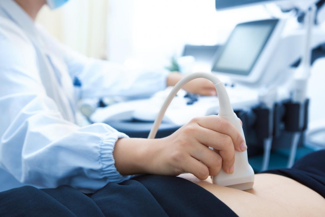
The ultrasound procedure will vary depending on the part of the body being scanned and potential health conditions. Usually, it is performed outside the body where you need to lie down on a table either on your side or your back. It does not require hospitalization and is safe and painless.
Once you are prepared, our highly experienced radiologist will apply a water-soluble gel to the body part being scanned. The gel allows the scanner (transducer) to smoothly move on your skin and capture high-quality images. The radiologist will then glide the transducer on your skin and begin scanning, gently pressing at some points and capturing images of different internal structures.
During the procedure, you need to stay still to ensure the images can be taken accurately. When the test is complete, wipe off the gel using tissue paper and you can get back to your daily life. The procedure does not require any recovery time.
In addition to the above ultrasound tests, we also specialize in:
- Transvaginal ultrasound where a special type of transducer is inserted into the vagina
- Transrectal ultrasound of the prostate that requires inserting the transducer through the rectum
- Transesophageal ultrasound of the heart in which the transducer is inserted into the esophagus, usually under sedation
A strong magnetic field is made by passing an electric current through the wire loop. Although this is happening, other coils send and receive radio waves in the magnet. It triggers the proton in the body to align itself. Once aligned, radio waves are absorbed by protons, that stimulate spinning. Energy is released after “exciting” the molecules, which in turn emits the energy signals raised by the coil. This information is then sent to the computer which processes all the received signals and generates them into an image. The final product is represented by the 3-D image of the field being examined.
We perform a wide range of procedures that include:
- General ultrasound
- Abdominal ultrasound
- Musculoskeletal ultrasound
- Breast ultrasound
- Prostate ultrasound
- Thyroid ultrasound
- Pregnancy ultrasound
For the above diagnostic imaging procedures, we use the latest 3D and 4D ultrasound technology for more precise image quality. Trust us for high-quality and reliable 3D ultrasound near me that provides three-dimensional images of the body’s internal structure or the fetus. The image quality is more accurate and detailed – aiding in better diagnosis.
We also specialize in 4D ultrasound near me in Delhi, taking medical imaging a step further. Through cutting-edge 4D scanning, we generate real-time, live images which are presented in the form of moving images or videos. Such imaging procedures are best performed to view the heart’s blood flow, diagnose fetal defects or experience the heartbeat of the unborn baby.
