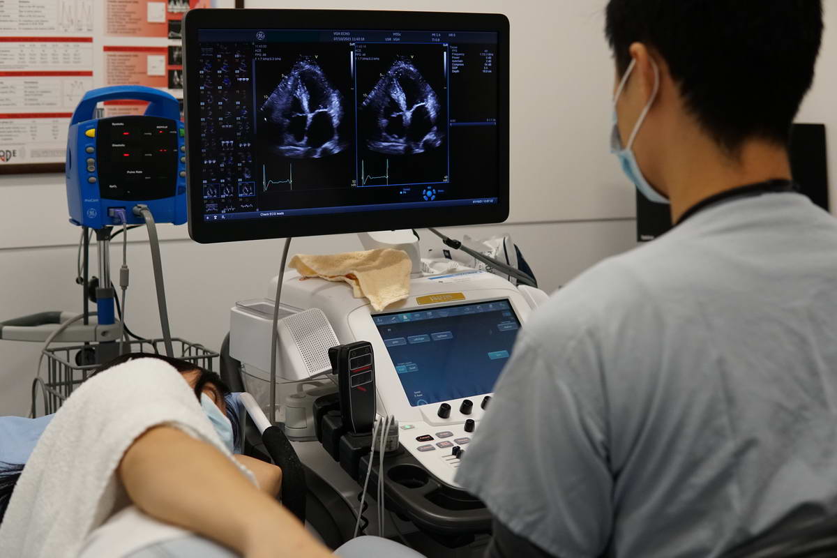
Echocardiogram
An echocardiogram is an ultrasound test that checks the structure and function of your heart. An echo can diagnose a range of conditions including cardiomyopathy and valve disease. There are several types of echo tests, including transthoracic and transesophageal.
An echocardiogram (echo) is a graphic outline of your heart’s movement. During an echo test, your healthcare provider uses ultrasound (high-frequency sound waves) from a hand-held wand placed on your chest to take pictures of your heart’s valves and chambers. This helps the provider evaluate the pumping action of your heart.
Providers often combine echo with Doppler ultrasound and color Doppler techniques to evaluate blood flow across your heart’s valves.
Echocardiography uses no radiation. This makes an echo different from other tests like X-rays and CT scans that use small amounts of radiation.
A technician called a cardiac sonographer performs your echo. They’re trained in performing echo tests and using the most current technology. They’re prepared to work in a variety of settings including hospital rooms and catheterization labs.
There are several types of echocardiogram. Each one offers unique benefits in diagnosing and managing heart disease.
Several techniques can be used to create pictures of your heart. The best technique depends on your specific condition and what your provider needs to see. These techniques include:
- Two-dimensional (2D) ultrasound. This approach is used most often. It produces 2D images that appear as “slices” on the computer screen. Traditionally, these slices could be “stacked” to build a 3D structure.
- Three-dimensional (3D) ultrasound. Advances in technology have made 3D imaging more efficient and useful. New 3D techniques show different aspects of your heart, including how well it pumps blood, with greater accuracy. Using 3D also allows your sonographer to see parts of your heart from different angles.
- Doppler ultrasound. This technique shows how fast your blood flows, and also in what direction.
- Color Doppler ultrasound. This technique also shows your blood flow, but it uses different colors to highlight the different directions of flow.
- Strain imaging. This approach shows changes in how your heart muscle moves. It can catch early signs of some heart disease.
- Contrast imaging. Your provider injects a substance called a contrast agent into one of your veins. The substance is visible in the images and can help show details of your heart. Some people experience an allergic reaction to the contrast agent, but reactions are usually mild.
An echocardiogram usually takes 40 to 60 minutes. A transesophageal echo may take up to 90 minutes.
An echocardiogram and an electrocardiogram (called an EKG or ECG) both check your heart. But they check for different things and produce different types of visuals.
An echo checks the overall structure and function of your heart. It produces moving pictures of your heart.
An EKG checks your heart’s electrical activity. It produces a graph, rather than pictures of your heart. The lines on this graph show your heart rate and rhythm.
Your provider will order an echo for many reasons. You may need an echocardiogram if:
- You have symptoms, and your healthcare provider wants to learn more (either by diagnosing a problem or ruling out possible causes).
- Your provider thinks you have some form of heart disease. The echo is used to diagnose the specific problem and learn more about it.
- Your provider wants to check on a condition you’ve already been diagnosed with. For example, some people with valve disease need echo tests on a regular basis.
- You’re preparing for a surgery or procedure.
- Your provider wants to check the outcome of a surgery or procedure.
An echocardiogram can detect many different types of heart disease. These include:
- Congenital heart disease, which you’re born with.
- which affects your heart muscle.
- which is an infection in your heart’s chambers or valves.
- which affects the two-layered sac that covers the outer surface of your heart.
- which affects the “doors” that connect the chambers of your heart.
An echo can also show changes in your heart that could indicate:
- Blood clots.
- A cardiac tumor.
