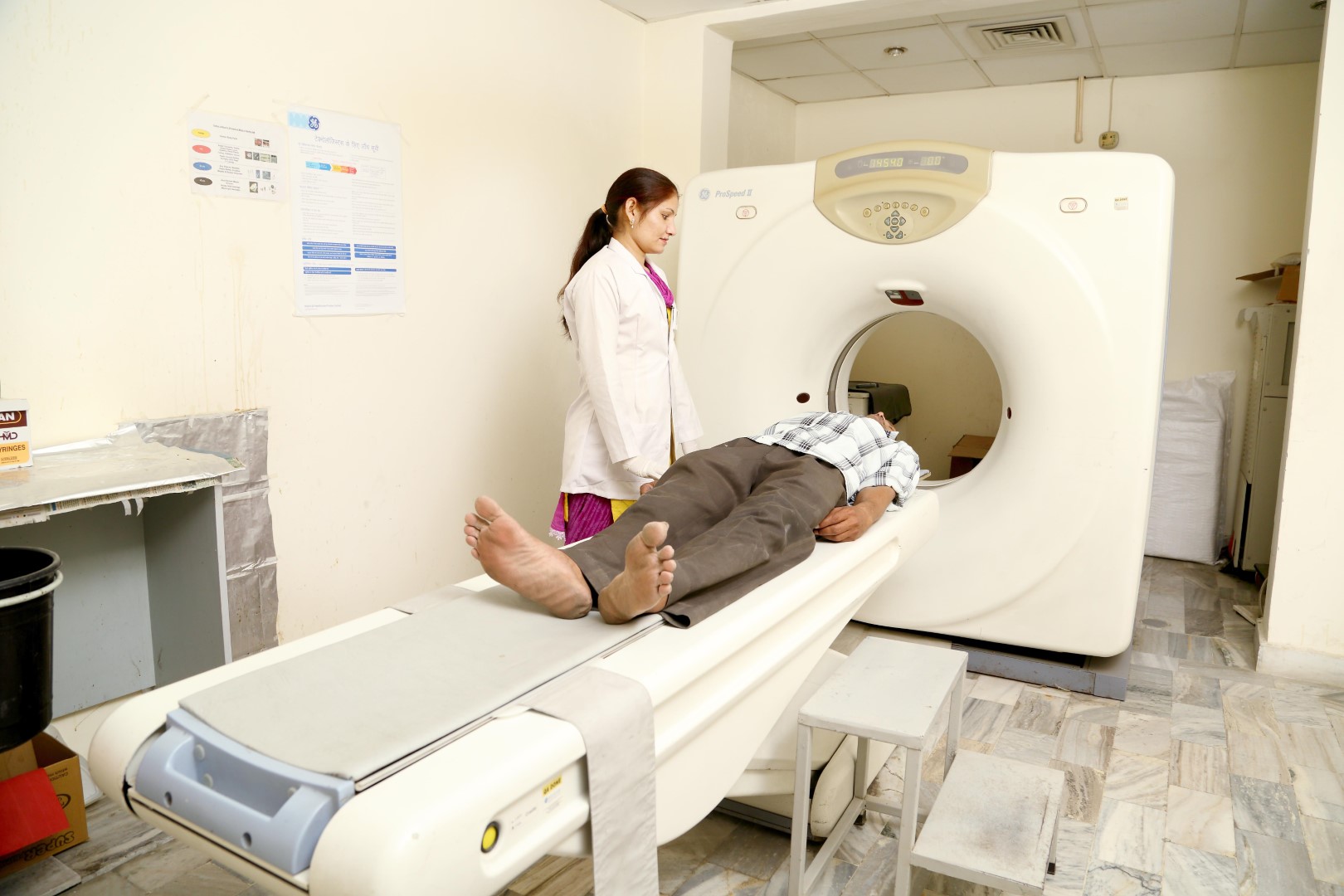
CT-Scan
Computed tomography (CT) is a diagnostic procedure that uses special X-ray equipment to create cross-sectional pictures of your body. CT images are produced using X-ray technology and powerful computers. The uses of CT include looking for Broken bones Cancer Blood clots Signs of heart disease Internal bleeding During a CT scan, you lie still on a table. The table slowly passes through the center of a large X-ray machine. The test is painless. During some tests, you receive a contrast dye, which makes parts of your body show up better in the image.
There are certain health conditions that need a deeper look into your body’s internal structures, including blood vessels, tissues and bones. Regular x-rays or ultrasound procedures may not always provide a detailed view. Hence, your doctor may recommend having a computed tomography (CT) scan done for a better diagnosis.
A CT scan procedure combines radiology imaging and computer technology to create a series of highly precise and cross-sectional images captured from different angles. All the pictures are combined to create a two-dimensional or three-dimensional image of a specific part of the body.
A CT scan can be used to have a better and more detailed view of all body parts and is primarily used to detect an injury or disease or to guide a medical, radiation or surgical treatment.
A CT scan can be used for a variety of reasons as mentioned herewith:
- Diagnose and monitor a variety of health conditions and diseases such as heart diseases, liver masses, lung nodules and other cancer
- Detect bone and muscle disorders such as fractures and bone tumours
- Identify the cause of internal bleeding or internal injuries
- Assess how a certain treatment is working, such as radiation for cancer treatment
- Guide specific procedures such as radiation therapy, biopsy and surgery
- Identify the specific location of a blood clot, infection or tumour
- Prepare for a certain treatment or surgery
When you arrive at a CT scan centre. you will require changing into a gown. During the scan, you need to lie down on a long narrow table. When you are ready, the table is slid inside a doughnut-shaped, circular CT scanner.
The multi-detector scanner has excellent image capabilities, creating high-resolution 2D and 3D images. The device has X-Ray beams that will circle the part of the body being scanned and capture cross-sectional images. The pictures are then digitally scanned on a computer system and processed to render a series of high-quality 2D and 3D representations of the body’s internal structure.
This is the standard procedure of most CT scan procedures. However, some complex tests require injecting a contrast material before the test. This helps achieve more detailed and accurate images of the part of the body being scanned.
