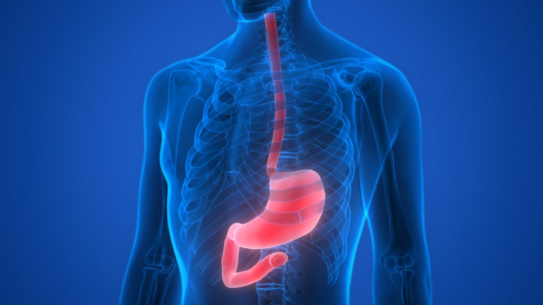
Barium X-Rays
These studies are specialized X-ray procedures using a solution containing barium to examine the gastrointestinal tract, namely the oesophagus, stomach, small intestine, and large intestine.
The most common studies are barium swallows, barium meals, and barium enemas.
A barium X-ray is a radiographic (X-ray) examination of the gastrointestinal (GI) tract. Barium X-rays (also called upper and lower GI series) are used to diagnose abnormalities of the GI tract, such as tumors, ulcers and other inflammatory conditions, polyps, hernias, and strictures.
The use of barium with standard X-rays contributes to the visibility of various characteristics of the GI tract. Barium is a dry, white, chalky powder that is mixed with water to make barium liquid. Barium is an X-ray absorber and appears white on X-ray film. When instilled into the GI tract, barium coats the inside wall of the esophagus, stomach, large intestine, and/or small intestine so that the inside wall lining, size, shape, contour, and patency (openness) are visible on X-ray. This process shows differences that might not be seen on standard X-rays. Barium is used only for diagnostic studies of the GI tract.
In addition to drinking barium, air is often inserted into the bowel for a lower GI X-ray. For an upper GI X-ray, some patients may be given baking soda crystals (similar to Alka-Seltzer) to further improve the image. These types of procedure are called air-contrast or double-contrast GI studies.
Fluoroscopy is often used during a barium X-ray. Fluoroscopy is a study of moving body structures—similar to an X-ray “movie.” A continuous X-ray beam is passed through the body part being examined, and is transmitted to a TV-like monitor so that the body part and its motion can be seen in detail. In a barium X-ray, fluoroscopy allows the radiologist to see the movement of the barium through the GI tract as it is instilled through the mouth or the rectum.
Reasons for performing barium X-ray procedures may include the following:
- Abdominal pain
- Bleeding from the rectum
- Unexplained vomiting
- Bowel movement changes
- Chronic diarrhea or constipation
- Pain or difficulty swallowing
- Unexplained weight loss
- Unusual bloating
- To detect anatomical abnormalities
- Additional procedures are often performed in addition to barium X-rays. These procedures may include endoscopic examinations (an endoscope is a thin, flexible tube that is inserted into a body cavity and, using fiberoptic technology, provides direct visualization of the inside of the cavity), computerized tomography (CT) or magnetic resonance imaging (MRI) scans, and intra-cavity ultrasound.
There are three types of barium X-ray procedures:
- Barium enema (also called lower GI series)
- Barium small-bowel follow through
- Barium swallow (also called upper GI series)
A barium enema involves filling the large intestine with diluted barium liquid while X-ray images are being taken. Barium enemas are used to diagnose disorders of the large intestine and rectum. These disorders may include colonic tumors, polyps, diverticula, and anatomical abnormalities.
Usually, a barium enema can be performed on an outpatient basis. The patient may be asked to do the following in preparation for a barium enema:
- Drink clear liquids the day before the examination.
- Follow a special liquid diet one to two days prior to the procedure.
- Take a laxative, suppository, or drug to cleanse the bowel.
- Refrain from eating and drinking after midnight on the night before the examination.
These measures are done to empty the large intestine, as any residue (feces) can obscure the image. However, a barium enema may be done without preparation, for example, to diagnose Hirschsprung’s disease.
Barium enemas are performed in two ways:
- Single-contrast image. The entire large intestine is filled with barium liquid. Single-contrast images show prominent abnormalities or large masses in the large intestine.
- Double-contrast image. A smaller quantity of thicker barium liquid is introduced to the large intestine, followed by air. Double-contrast images show smaller surface abnormalities of the large intestine, as the air prevents the barium from filling the intestine. Instead, the barium forms a film on the inner surface.
Although each hospital may have specific protocols in place, generally, a barium enema procedure follows this process:
- The patient will be positioned on an examination table.
- A rectal tube will be inserted into the rectum to allow the barium to flow into the intestine.
- The radiologist will use a machine called a fluoroscope (a device used for the immediate showing of an X-ray image).
- During the procedure, the machine and examination table will move and the patient may be asked to change positions.
- Additional X-rays may be made immediately after the procedure in order to obtain greater details of the area under examination. Often, additional X-rays are made after the barium has been excreted from the bowel, which is usually one or more days after the procedure.
- After the procedure, a small amount of barium will be expelled from the body immediately. The remainder of the liquid is later excreted in the stool. Barium liquid may cause constipation and light colored stools. Following the examination, the patient may be asked to eat foods high in fiber and drink plenty of fluids to help expel the barium from the body. If you do not have a bowel movement for more than two days after your exam or are unable to pass gas rectally, call your physician promptly. You may need an enema or laxative to assist in eliminating the barium.
A barium small-bowel follow through involves filling the small intestine with barium liquid while X-ray images are being taken. Barium small-bowel follow throughs are used to diagnose disorders of the small intestine, such as ulcers, tumors, and inflammatory bowel disease, a group of disorders that includes Crohn’s disease and ulcerative colitis.
Usually, a barium small-bowel follow through can be performed on an outpatient basis. Patients may be asked to refrain from eating or drinking after midnight on the night before the examination. An enema or laxative may be given on the day before the test to clear feces from the bowel.
Although each hospital may have specific protocols in place, generally, a barium small-bowel procedure follows this process:
- the patient is given a bottle of barium to drink.
- The patient is positioned on the examination table.
- The radiologist uses a machine called a fluoroscope (a device used for the immediate showing of an X-ray image).
- X-rays are taken every 20 to 30 minutes over the next hour or two until the entire small bowel is opacified. This exam can take several hours
- Following the examination, the patient may be asked to eat foods high in fiber and drink plenty of fluids to expel the barium from the body. If you do not have a bowel movement for more than two days after your exam or are unable to pass gas rectally, call your physician promptly. You may need an enema or laxative to assist in eliminating the barium.
An upper GI series is an examination of the esophagus and stomach using barium to coat the walls of the upper digestive tract so that it may be examined under X-ray. An upper GI that focuses on the esophagus is also known as a barium swallow. Barium swallows and upper GI series are used to identify any abnormalities such as tumors, ulcers, hernias, pouches, strictures, and swallowing difficulties.
Usually, these tests can be performed on an outpatient basis. Patients may be advised not to eat or drink after midnight on the night before the examination.
Although each hospital may have specific protocols in place, generally, the procedure follows this process:
- The patient will be asked to drink the barium liquid and to swallow baking soda crystals. It is important not to belch, as the gas assists the radiologist in evaluation.
- The patient will stand behind a machine called a fluoroscope (a device used for the immediate showing of an X-ray image).
- The patient may be asked to move in different positions and to hold his or her breath while the X-rays are taken.
- If the small intestine is to be examined, the patient may be asked to drink additional barium and a series of X-rays will be taken until the barium reaches the colon.
- Following the examination, barium may cause constipation. The patient may be advised to drink plenty of fluids and eat foods high in fiber to expel the barium from the body. If you do not have a bowel movement for more than two days after your exam or are unable to pass gas rectally, call your physician promptly. You may need an enema or laxative to assist in eliminating the barium.
