CBCT
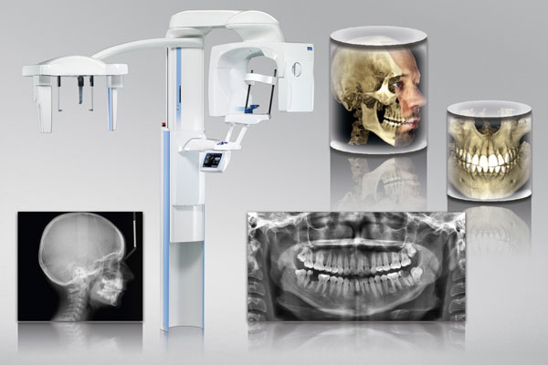
The Cone Beam CT uses an amorphous silicon flat panel detector and cone beam technology to get volumetric images with vastly lower radiation dose and higher precision when compared to traditional 16 or 64 slice Ct Scanner. This new age device provides a full range of diagnostic views including complete scans of all oral and maxillofacial structures, paranasal sinuses and airways. Acquired information gives the treating doctor more thorough structural knowledge which enables highly accurate diagnosis and treatment planning and can help create more predictable outcomes. Vastly lower radiation dose (20-100 times) means that the CBCT scan can be done safely without any undue concern for radiation side-effects.
Computed tomography has been expanding rapidly and CBCT is the latest, most advanced generation of CTs. Cone Beam technology uses a cone shaped X-Ray beam that projects onto an amorphous silicon flat panel detector. The scanner rotates 360 degrees around a patient’s head in a matter of seconds with the patient in a comfortable sitting position. The single turn motion image capture used in Cone Beam CT is quicker than conventional spiral motion, and can be accomplished at a lower radiation dose as a result of no overlap of slices. Cone beam CT provides views that can be presented as 3D volumes or 2D images for diagnosis and advanced planning.
Traditional multi slice (6, 8, 16,32,40,64,128,256 and 320) CT scanners have been used in the past for dental and maxillofacial imaging.
Very Low Radiation Dose: The CBCT gives a radiation dose of about 36~Sv as compared to 2000~Sv to 4000~Sv of multi-slice CT.
Higher Precision and Accuracy: The CBCT provides images of superb clarity and great precision. What you see is real and the measurements are very accurate.
Dedicated Scanner: The CBCT is a dedicated scanner designed specifically for dental and maxillofacial imaging.
Dedicated Software: The dedicated user-friendly software simplifies treatment planning and provides necessary perspective in many cases. True-size, distortion free, high-resolution images are reconstructed rapidly, tailor-made for the needs of dental and maxillofacial professionals.
Eliminates Need for Multiple Exposures: The CBCT provides all information in one scan including panoramic and cephalometric images and 3D volumes, virtually eliminating the need for conventional orthodontic X-Rays. Patients can be comforted by the knowledge that the doctor will have all the information needed to evaluate their problem and plan treatment.
- Impaction/ Localization of teeth or Foreign body
- Oral surgery/Third Molar inerve relationship
- Orthodontics
- Pre-Implant imaging/Placement/Planning
- Intra-bony pathology
- TMJ Studies
- Asymmetry studies
- Orthognathic surgery
- Cosmetic surgical planning
- Endodontic canal assessment
- Airway evaluation
- Para nasal sinus investigation